Mineral Services for Mining Exploration and Production
DCMSL uses the following tools for mineral/material analysis:
X-ray Diffraction – X-ray diffraction (XRD) is a tool for the investigation and identification of minerals and materials by their atomic structure. This technique is highly useful in the determination of whole rock mineralogy and single phase analysis.
Light Microscopy – Analytical microscopy draws on techniques used in the fields of geology, chemistry, biology and forensics. Microscopy provides rapid, non-destructive analysis and requires very little material. Optical properties of materials/minerals in either transmitted light or reflected light are identified and may be compared to the properties of known standards.
Scanning Electron Microscopy – The scanning electron microscope (SEM) has unique capabilities for analyzing surfaces and forms an image using electrons. The SEM provides high resolution, extensive ranges of magnification and a high depth of field. Equipped with an energy dispersive spectrometer (EDS), chemical information of individual grains can be determined.
Industrial Minerals
Over the past 30+ years DCMSL has worked on numerous industrial mineral projects. Most of these projects have centered around product purity, contamination and physical separation characteristics. Our experience includes:
Industrial Silicates
- Determine aspect ratio of wollastonite and talc
- Determine trace impurities and inclusions
- Determine free crystalline silica content
- Determination of asbestiform minerals
Heavy Mineral Sands
- Determine ratios of ilmenite, rutile/anatase and leucoxene
- Determine amounts of zircon, monazite and other trace minerals
- Evaluate free crystalline silica
Silica Sands
- Determine identity of opaque inclusions that may impact total Fe content
- Utilize heavy mineral separations to determine type and quantity of refractory mineral phases.
X-Ray Diffraction Analysis
(XRD) is used to identify a wide variety of crystalline compounds from pharmaceuticals, bulk mineral assemblages in ores, whole rock, clays and metallurgical concentrates or metallic powders.
DCMSL has two Phillips x-ray diffraction units equipped with four goniometers capable of performing line scans for qualitative work and step scanning for semi-quantitative analysis using standard powder packs or zero background plates for sample sizes as small as a few milligrams. Computer controlled scans are processed utilizing Jade 9 software using a search match function to access a JCPDS database. This database of over 35,000 inorganic and organic materials is available to match any unknown phase with a standard.
Depending on the needs of the client several XRD analysis options are available:
Qualitative XRD Scan
Since all crystalline material produces a unique X-ray diffraction pattern, XRD is a powerful tool for the qualitative identification of unknown single phases or mixed phases. Identification is made using search match software to access a JCPDS database of reference patterns.
Semi-Quantitative XRD Analysis
Estimates of mineral concentrations in multicomponent samples with moderate accuracy is achieved using the Reference Intensity Method (RIR). Phase concentration is based on peak height/area and RIR’s measured in house or from published data.
<2µm Clay Speciation and Semi-Quantitative XRD Analysis
Oriented clay mounts (<2μm) are prepared using a peel method and analyzed glycolated, air dried and heated for clay identification. Concentrations of individual clay species are based on integrated peak areas and intensity factors measured in house on known standards or computer calculated.
Optical Microscopy
DCMSL considers the light microscope to be one of the most important basic tools in the laboratory. While most companies concentrate on the big picture, DCM Science Laboratory puts their focus on the little things… microscopic materials magnified 10 to 1000 times. DCMSL has a wide variety of microscopes and accessories to solve problems or provide standard analyses for a wide variety of industries.
- Stereo binocular microscope. A low power microscope (6 to 80X) used for sample observation, manipulation, and preparation. Lighting systems include inclined incident, coaxial incident and fiber optic illuminators.
- Phase contrast microscope. A transmitted light microscope (40 to 1000X) used for observation and identification of structures in materials with very low relief.• Polarized light microscope. A transmitted light microscope (40 to 1000X) used for observation and identification of non-opaque materials.
- Reflected light microscope. An incident light microscope (50 to 1000X) used for observation and identification of opaque materials.
- Photography. Utilizing highly specialized photographic equipment, DCMSL can meet any analytical need in the areas of photomicrography. The Moticam 2500 digital system adapts to any microscope, (stereo binocular, polarized light, fluorescent, and others) using digital formats for reports or professional papers. The DCMSL photography staff provides fast, professional results tailored to specific client needs and budgets.
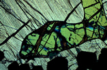
Petrography
Petrography is the study of rocks in specially prepared thin section by both reflected and transmitted light microscopy and is the most effective way to classify rock types. The study of opaque ore minerals in polished sections is one of the most widely used techniques for identification of ore phases and their textural relationships with gangue minerals. DCMSL has 35+ years of experience in analytical petrography and can customize an analysis that best achieves our clients’ objectives. For example, we can provide:
- Basic rock description with identification of major rock forming minerals and rock type. Comments on deleterious phases that may react with cement for aggregate clients.
- Ore petrography that includes identification of opaque phases, gangue minerals, alteration products, rock type and paragenesis where reasonably obvious. Grain size and locking characteristics of ore mineral with gangue are also described.
- Digital photomicrographs are included with all petrographic studies that highlight relevant aspects.
- Petrographic evaluation of concentrates and tailings to help solve problems encountered in mineral processing. Petrographic studies of this type are used to determine:
- Particle size and textural association with gangue.
- Liberation characteristics in feed and final products
- The amount of residual target minerals in the tailings or gangue material in final concentrates.
Scanning Electron Microscopy
A scanning electron microscope (SEM) generates a three-dimensional image of objects by directing a finely defined electron beam on the surface of a specimen. The surface topography is displayed as an image by electrons and energy ejected from the surface. The use of electrons, as opposed to visible light, allows high resolution combined with a great depth of field and high magnification of a material’s surface. The range of magnification (10 to 500,000X) overlaps the light microscope at the low end and the transmission electron microscope at the high end. Imaging is achieved using either secondary electrons or backscatter electrons (BSE). BSE imaging is particularly useful with the ore minerals. Since heavy elements backscatter electrons more strongly than light elements, compounds composed of heavy elements appear brighter in the image. BSE imaging is one of the most useful tools in locating sub-micron gold in sulfides or silicate gangue minerals otherwise impossible by secondary imaging. Coupled with an energy dispersive spectrometer (EDS) chemical information in grains as small as 1µm can be achieved. EDS detects all elements except H, He, Li and Be. EDS can be used for quantitative analysis and x-ray mapping. DCMSL has utilized SEM/EDS services for our clients for over 30 years and can customize an analysis to meet their needs.
The powerful combination of the SEM with the EDS x-ray microanalysis system has broad applications in the following areas:
Qualitative Elemental Analysis
Microanalysis Quantification
Semi-quantitative Analysis
X-ray Mapping
Grain Boundary Reconstruction
Grain Sizing
Comparative Studies
Microstructural Analysis
Mineralogy

Quality control in the laboratory is maintained by a rigid, constantly-monitored quality assurance program.
DCMSL is accredited by the American Industrial Hygiene Association (AIHA LAP, LLC) since 1986 and by the National Voluntary Laboratory Accreditation Program (NVLAP Lab Code 101258-0) since 1989.